Plant cell diagram pencil sketch simple functions and diagram
Table of Contents
Table of Contents
If you’re a student or a teacher looking for a fun and creative way to teach animal cells, drawing your own 3D animal cell is a great way to learn about the different parts of the cell. But, drawing a 3D animal cell can be challenging and time-consuming. In this article, we will give you tips and tricks on how to draw a 3D animal cell quickly and easily.
When it comes to drawing a 3D animal cell, some common pain points are knowing where to start and what each part of the cell looks like. Without a clear understanding of what an animal cell looks like, it can be difficult to know where to start drawing or what each part of the cell does.
First, let’s answer the question of how to draw a 3D animal cell. To get started, you’ll need a few materials: a pencil, paper, a ruler, and colored pencils or markers if you want to make your cell vibrant and colorful. Next, study a few reference images of an animal cell to fully understand the different parts of the cell and how they fit together. Once you have a good understanding of the different parts of the cell, draw an oval shape to represent the cell’s membrane. Then, add in the different organelles and label them accordingly with colored pencils or markers.
To summarize, drawing a 3D animal cell involves understanding different parts of the cell and creating the cell’s organelles on paper. By following these tips and tricks, you can easily draw a 3D animal cell in no time.
How to Draw a 3D Animal Cell: Tips and Tricks
As someone who has struggled with drawing 3D animal cells in the past, I found that it’s helpful to break down each part of the cell into its own drawing. This helps me make sure that each part of the cell is accurately depicted before adding them to the cell as a whole. Additionally, it’s important to not get bogged down in the details. While it’s important to represent the organelles accurately, you don’t need to be a skilled artist to draw a good 3D animal cell.
Understaning the Parts of an Animal Cell
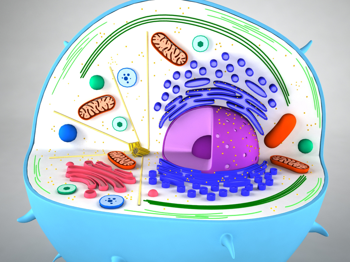
The parts of an animal cell include the nucleus, mitochondria, endoplasmic reticulum, ribosomes, golgi apparatus, lysosomes, and centrosomes. The nucleus is considered the cell’s control center, while the mitochondria is responsible for producing energy in the cell. The endoplasmic reticulum is where proteins are synthesized and modified, while the ribosomes are the site of protein synthesis. The golgi apparatus is responsible for processing and packaging substances produced by the cell, while lysosomes are involved in breaking down waste materials. Finally, the centrosomes play a role in cell division.
Different Shapes and Sizes
It’s important to note that some organelles in an animal cell come in different shapes and sizes. For instance, lysosomes can vary in size and shape depending on what they’re breaking down. Don’t worry too much about getting the shape and size of each organelle perfectly accurate. The goal is to provide a basic representation of each organelle and its location in the cell.
Adding Color to Your 3D Animal Cell
If you want to make your 3D animal cell stand out, consider adding colors to the different organelles. While it’s important to not get too bogged down in the details, adding a colorful touch can help make your cell more visually appealing. The nucleus is usually depicted in blue, while the mitochondria takes on a reddish-pink hue. The endoplasmic reticulum and ribosomes are usually drawn in yellow, while the golgi apparatus is shaded with a touch of purple.
Question and Answer
Q1. Is it necessary to make a 3D animal cell for science fair projects?
A. While making a 3D cell is not necessary for science fair projects, it is a great way to visually represent an animal cell and its different organelles.
Q2. Can beginners draw a 3D animal cell?
A. Yes, beginners can draw a 3D animal cell by studying reference images of animal cells and breaking down each part of the cell into its own drawing.
Q3. What colors can be used to represent the organelles of an animal cell?
A. The nucleus is usually depicted in blue, while the mitochondria takes on a reddish-pink hue. The endoplasmic reticulum and ribosomes are usually drawn in yellow, while the golgi apparatus is shaded with a touch of purple.
Q4. Is it important to label each part of the animal cell?
A. Yes, it’s important to label each part of the animal cell to help understand the function of each organelle.
Conclusion of how to draw a 3D animal cell
By following the tips and tricks provided in this article, drawing a 3D animal cell can be quick and easy, even for beginners. Don’t worry too much about getting each organelle’s shape or size perfectly accurate. Simply aim to provide a basic representation of each organelle and its location in the cell. Adding colors to your drawing can also help make your cell more visually appealing.
Gallery
4 Ways To Make An Animal Cell For A Science Project | Science Projects

Photo Credit by: bing.com /
Plant Cell Diagram Pencil Sketch Simple : Functions And Diagram

Photo Credit by: bing.com / drawing animals
How To Draw Animal Cell Step By Step Tutorial For Beginners ! - YouTube

Photo Credit by: bing.com / cell animal drawing draw sketch simple diagram plant label paintingvalley labeled step tutorial functions drawings parts explore
Animal Cell 3D Model - 3D Models World

Photo Credit by: bing.com / cell animal 3d model models
Animal Cell Model Labeled - Bing Images | Teaching | Pinterest | Cell

Photo Credit by: bing.com / styrofoam labeled


